For spectral cytometry, we need to extract the spectra of the fluorochromes used one by one.
To this end, it will be necessary to provide:
1) Completely unstained cells. The autofluorescence of cells is indeed considered as a fluorochrome. It is therefore possible to evaluate the autofluorescence spectrum and subtract it from the fluorescence signals.
2) All single stained (tubes containing cells stained with only one fluorochrome. The preparation of single stained cells must be the same of the sample (dissociation, washing, permeabilization, fixation, etc.). If you are not sure about the presence of a marker on your cells (or you have not enough cells for all controls) and in order to extract the spectrum, you will need to provide beads that bind your antibodies. The beads must undergo the same treatments as that of cells. You can also find beads for viability markers.
Be careful : Avoid using beads for tandem fluorochromes (for instance : PE-Cy5.5, APC-Cy7, BV570, QDOT655..) because the extraction of the spectra is not done correctly when applied to cells :

Be careful : For unlabeled control we need minimum 100 000 cells (in about 200-300 µl) and for positive controls we need minimum 200 positive cells in order to extract the spectra.
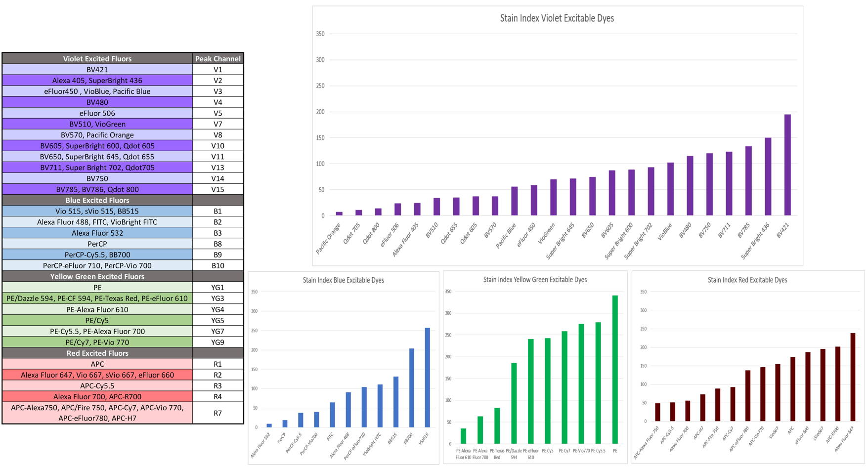
How to choose fluorochromes
https://welcome.cytekbio.com/hubfs/Website%20Downloadable%20Content/Data%20Sheets/Fluorochrome%20Guides/N9_20018_Rev._A_4L_VBYGR_Fluor_Guide.pdf
-the excitation wavelengths of the fluorochromes correspond to the light sources of the cytometer,
-the emission wavelengths are far enough apart that their signals can be analyzed separately.
-lowly expressed antigens are revealed by high yield fluorochromes and highly expressed antigens with low yield fluorochromes.
-in the case of co-expression on a cell, use fluorochromes with little or no overlapping spectra.
Elimination of dead cells
The elimination of dead cells can be done on the basis of the physical parameters side scatter (SSC) and forward scatter (FSC) for cell line samples. The addition of a dead cell marker (such as propidium iodide, DAPI, Sytox, etc.) allows more precise elimination for more complex samples. If a viability marker is used, the negative control (unstained) must not contain the marker.
Cell filtration
Cell filtration, just before analysis by the cell sorter, allows avoiding blockings that destabilize cell sorting and decrease sorted fractions purity. To this you can use 40 micron filters.
Conditioning cells before sorting
The cells can be brought in PBS or culture medium. The cells must be brought in 5ml tubes (reference Falcon 2054) with a minimum volume of 300 µl at:
• 5x10⁶ cells/ml maximum for low pressure sorting
• 1x10⁷ cells/ml maximum for intermediate pressure sorting
• 2 to 5x10⁷ cells/ml maximum for high pressure sorting
Be careful :The larger the volume to pass, the longer the sort will take.
Collection tubes
The cells can be collected after sorting in tubes (1.5ml Eppendorf tubes, 5ml FACS tubes, 15ml Falcon tubes) or plates (96, 24,12 and 6 wells). It is advisable to put culture medium in the collection tubes.
With the 100µ nozzle we can get about :
• 1x10⁶ cells in a 5ml tube
• 3x10⁶ cells in a 15ml tube
with the 70µ nozzle :
• 3x10⁶ cells in a 5ml tube
• 9x10⁶ cells in a 15ml tube
Adjust collect tubes number to the collected cells estimated number for each fraction.